
Fig. 1.
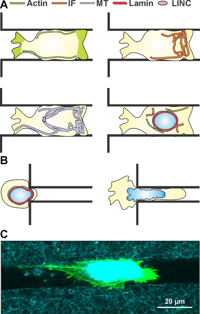
Overview of key mechanical features governing cell migration in restrictive 3-dimensional (3D) channels. A: representations of the same migrating cell inside a restrictive 3D channel showing cortical localization of actin along the wall of the channel and at both extremities of the migrating cell, intermediate filament (IF) localization at the leading edge of the migrating cell, microtubule (MT) preferential localization at the leading edge of the migrating cell, and a schematic of cytoskeletal-nucleoskeletal connections via linker of nucleoskeleton and cytoskeleton (LINC) complex coupling of lamins and actin/IFs. B: overexpression of lamin IFs stiffens the nucleus and poses a significant barrier to migration in subnuclear pore sizes, while diminished expression of lamin IFs softens the nucleus and allows for greater nuclear deformability. C: confocal image of a cell migrating inside a microfabricated 3D collagen microtrack showing actin (green) and reflectance (collagen, cyan). (See Ref. 78 for details.)