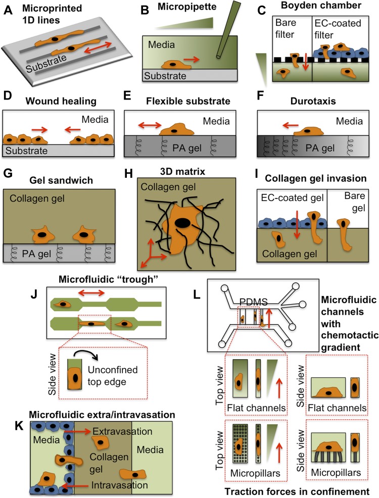
Fig. 2.
Diverse in vitro assays have been used to explore physical and biological cues affecting cell migration. Each of these assays mimics one or multiple cues presented to tumor cells (orange) and/or the endothelium (blue) during metastasis. Note that this is not an exhaustive list of migration assays. A: microphotopatterned or microcontact printed ECM protein lines (21, 33). B: micropipette chemotaxis assay (95). C: Boyden chamber chemotaxis assay, with bare filter containing nanometer- to micrometer-sized pores, or with EC-coated filter (56, 85). D: wound-healing assay. E: polyacrylamide (PA) gel flexible substrate assay (108). F: durotaxis assay using PA gel with a gradient of stiffness (87). G: collagen-PA gel sandwich assay (40). H: 3-dimensional (3D) collagen gel matrix assay (43). I: collagen gel invasion assay, with bare gel or EC-coated gel (93). J: microfluidic “trough” assay with 3 confining edges (106). K: microfluidic extra-/intravasation assay (70, 157). L: microfluidic chemotaxis assay with flat (5, 137) or micropillar-containing (114) channels. PDMS, polydimethylsiloxane. Red arrows indicate direction of cell migration.

