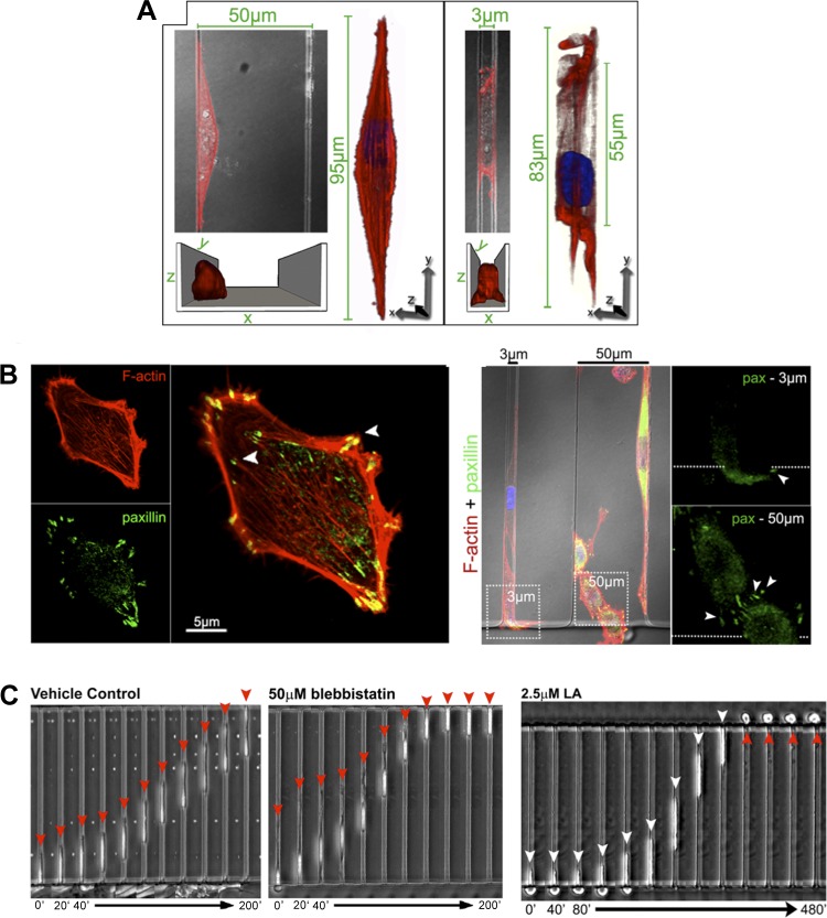
Fig. 3.
Physical confinement alters tumor cell adhesion and migration mechanisms. A: confocal reconstructions of the actin cytoskeleton (red; phalloidin staining) reveal significantly altered actin morphology in physically confined MDA-MB-231 metastatic breast tumor cells within narrow microchannels (3 μm wide × 10 μm high) compared with wide channels (50 μm wide × 10 μm high). B: confocal images of F-actin (red) and paxillin (green) on 2D planar surface (left) and in narrow and wide microchannels (right). Paxillin-positive punctate focal adhesions are much less pronounced in narrow than in wide channels. C: MDA-MB-231 cells migrate across narrow (3 μm wide × 10 μm high) channels, even in the absence of myosin II-mediated contractility (blebbistatin) or actin polymerization [latrunculin-A (LA)]. Time below each image series is in minutes. [From Balzer et al. (5).]

