Fig. 2.
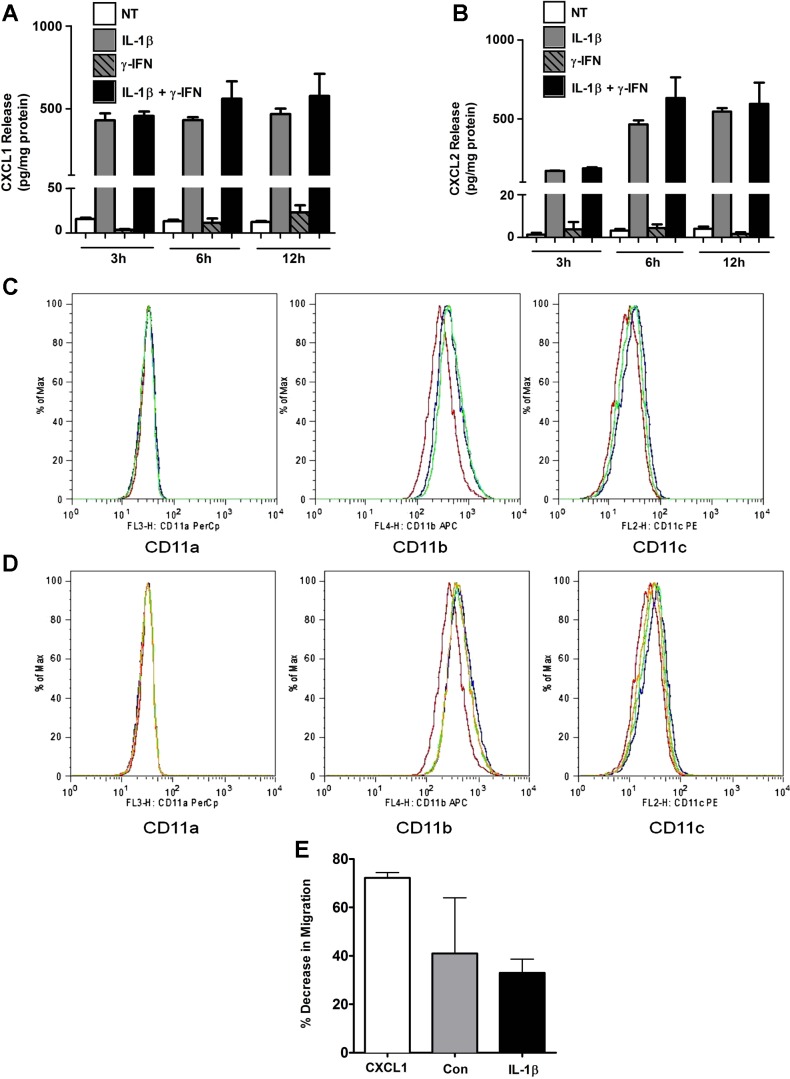
IL-1β promotes release of the CXCL1 and CXCL2 chemokines and activates peripheral blood neutrophils (PBN) for integrin expression and migration via CXCR2. A and B: 832/13 cells were untreated or treated with 1 ng/ml IL-1β, 100 U/ml IFNγ, or the combination of both cytokines for 0, 3, 6, and 12 h. CXCL1 (A) and CXCL2 (B) release into culture medium was measured by ELISA and normalized to protein content via BCA assay. Values are presented as means ± SE from 3 individual experiments. PBN were unstimulated (PBS control, red lines) or exposed to CXCL1 (blue) or CXCL2 (green) at 100 nM and the level of expression of intergrins CD11a, CD11b, or CD11c measured by flow cytometry (C). PBNs were unstimulated (red) or incubated with a combination of CXCL1/CXCL2 at (1 nM/10 nM, green lines) or CXCL1/CXCL2 at 10 nM/1 nM (blue lines), and levels of integrin expression were measured (D). Plots are representative experiments from 3 replicates. PBNs were exposed to human CXCL1 (10 nM) or 1:10 dilutions of supernatants from control 832/13 (Con) or IL-β exposed (IL-1β) in the presence of CXCR2 inhibitor SB225002 (400 nM) in a chemotaxis assay (E). Bars, are average decrease in migration in the presence of the CXCR2 inhibitor relative to control with no inhibitor. Each bar is the average of 4–6 duplicates ± SD from 6 biological replicates.

