Fig. 6.
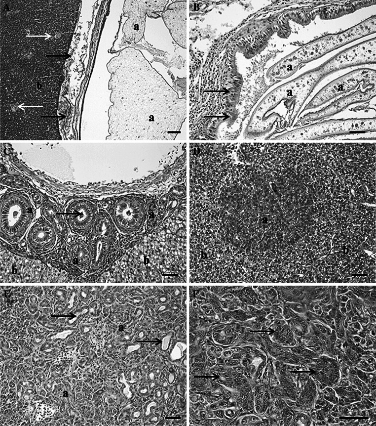
Microscopic pathology observed in the liver of white sucker collected within the St. Louis area of concern. a Encysted cestode (a) within the liver tissue (b) separated from hepatocytes by a layer of inflammatory and necrotic cells (black arrow). Macrophage aggregates (white arrows) were commonly observed within hepatic tissue. Scale bar 100 μm. b Sporoplasm of a myxozoan parasite (a) within a distended bile duct with areas of epithelial hyperplasia (black arrows). Scale bar 50 μm. c Focal area of bile duct proliferation (a) within hepatic tissue (b) with cross-sections of myxozoan sporoplasms (arrows). Scale bar 50 μm. d Foci of cellular alteration (a) within hepatic tissue (b). Scale bar 50 μm. e Section of a cholangiocarcinoma (a) illustrating well differentiated proliferating bile ductules containing bile (arrows). Scale bar pleomorphic cells, 50 μm. Hematoxylin and eosin stain

