Fig. 2.
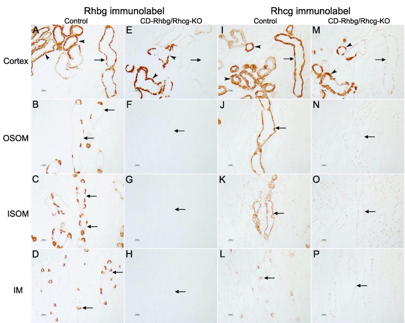
High-magnification immunohistochemical localization of Rhbg and Rhcg expression in C and CD-Rhbg/Rhcg-KO mice fed a normal diet. A and I: Rhbg and Rhcg immunolabel in C mice kidney in the cortex. A normal distribution of Rhbg and Rhcg immunolabel in the cortical collecting duct (CCD; arrows), in connecting tubule (CNT), and distal convoluted tubule (DCT; arrowheads) segments is present. E and M: Rhbg and Rhcg immunolabel in CD-Rhbg/Rhcg-KO mice kidney in the cortex. No Rhbg and Rhcg immunolabel is evident in CCD (arrows). Rhbg and Rhcg immunoreactivity is present in CNT and DCT segments (arrowheads). B–D and J–L: Rhbg and Rhcg immunolabel in OM and IM of C mice; normal Rhbg and Rhcg expression in the OSOM, ISOM, and IM (arrows). F–H and N–P: OM and IM from CD-Rhbg/Rhcg-KO mice kidney. No Rhbg or Rhcg immunolabel is detectable.

