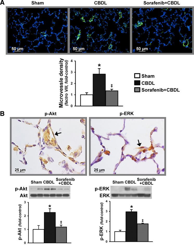
Fig. 5.
Pulmonary microvascular Akt and ERK phosphorylation after CBDL and effects of sorafenib on lung angiogenesis and RTK activation. A: representative immunofluorescence images of lung microvessel staining (factor VIII in green) and quantitation of microvessel density after CBDL. B: representative immunohistochemical staining images of p-Akt (Ser473) and p-ERK (Thr202/Tyr204) in the lung microvasculature (shown by arrows) after CBDL and representative immunoblots and graphical summaries of protein levels for p-Akt/Akt and p-ERK/ERK. Values are expressed as means ± SE (n = 8 animals for each group). *P < 0.05 compared with sham. ‡P < 0.05 compared with CBDL.

