Fig. 3.
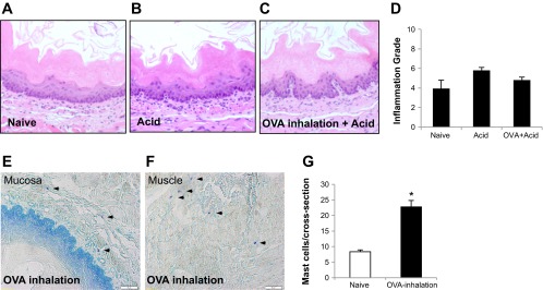
Histology of the esophagus: 1) in hematoxylin and eosin stain (A, B, C, and D), compared to control, intraluminal acid infusion in the esophagus from either naive or OVA-inhaled animals did not induce severe tissue damage (no erosion or ulceration) and only slightly increased inflammation grades (control vs. acid, or vs. OVA + acid = 3.89 ± 0.9 vs. 5.78 ± 0.3, or vs. 4.78 ± 0.3, P > 0.05, n = 4 in each group); 2) in toluidine blue stain (E, F, and G), compared with naive animals (n = 3), OVA inhalation significantly increased mast cell infiltrations (arrowheads) in the esophagus from OVA-sensitized animals (n = 4; naive vs. OVA inhalation: 8.33 ± 0.62 vs. 22.75 ± 2.07/cross-section, *P < 0.01).

