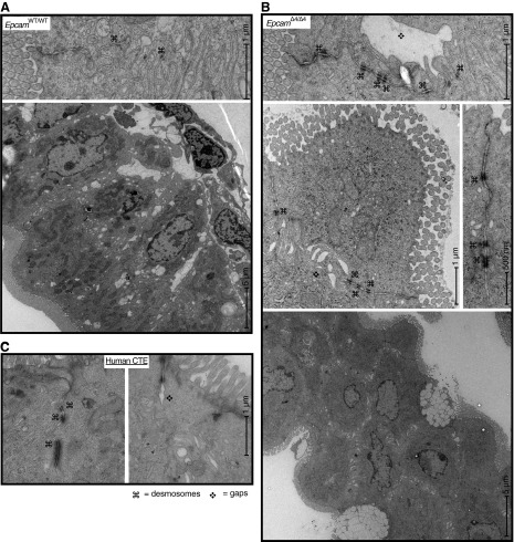
Fig. 3.
Electron microscopy of EpcamWT/WT (A) with normal ultrastructure compared with EpcamΔ4/Δ4 (B) mice showing intercellular gaps and increased length and number of desmosomes in EpcamΔ4/Δ4 mice. Gross structural changes, including loss of columnar shape (bottom right) were also seen. Images are representative of four EpcamΔ4/Δ4 mice and five littermate controls. C: electron microscopy of patients with CTE, demonstrating increased number and length of desmosomes (⌘) and intercellular gaps (❖).

