Fig. 4.
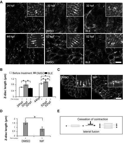
Contraction is required primarily for the lateral growth of Z-discs. A: the Z-discs in CMs revealed by α-actinin staining in 1-dpf (top) and 2-dpf (bottom) fish embryos, before and after the fish were treated with DMSO and 8 μmol/l BLE for 8 h. The 2 typical striated Z-dots or Z-discs in each group are indicated by arrows, whereas the randomly dispersed α-actinin dots, which do not assemble to myofibrils, are indicated by arrowheads. Original scale bar, 10 μm. B: Z-disc length, defined as the length of the Z-disc along the axis perpendicular to the myofibril, is increased during development but reduced significantly by BLE treatment. *P < 0.01. C: α-actinin staining in fish hearts for 2-dpf zebrafish embryos after the fish were treated by DMSO or 60 mM Nifedipine (NIF) for 8 h. The Z-disc becomes significantly shorter in Nifedipine-treated fish compared with DMSO-treated fish. Original scale bar, 20 μm. D: the length of the Z-disc is reduced dramatically by Nifedipine treatment. *P < 0.01. E: the schematic diagram illustrates the lateral fusion step in sarcomere assembly attenuated by cessation of contraction.

