Fig. 7.
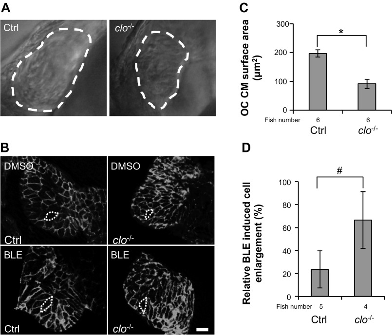
CM remodeling is independent of the endocardium. A: the ventricular chamber outlined by the white, dashed lines is smaller in cloche mutant fish than in control fish. clo−/−, cloche mutants. B: the outline of ventricular CMs was revealed by β-catenin staining in 2-dpf cloche mutant fish and control fish after the fish were treated with either DMSO or 8 μmol/l BLE for 8 h. The treatment started at 44 hpf, and heart samples were collected at 52 hpf. Original scale bar, 20 μm. C and D: quantification of cell size of CMs in B. BLE treatment elicits a higher degree of cell enlargement in cloche mutants than in control fish (D) despite the smaller cell size in cloche mutants than control fish in the DMSO treatment condition quantified in C. *P < 0.01; #P < 0.05.

