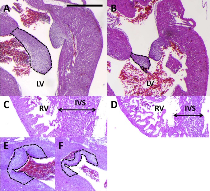
Fig. 1.
Late-stage congenital heart defects after ethanol exposure included smaller atrio-ventricular (AV) valves. A: control heart has large septal leaflet for left AV valve (outlined in black). Similar large AV leaflets were observed for all samples in this group (n = 4). LV, left ventricle. B: ethanol-exposed embryo developed a smaller AV septal leaflet (outlined in black). Small or malformed AV leaflets were observed for 5 of the embryos in this group (n = 7). C: control heart had a thick right ventricular (RV) wall and a thick interventricular septum (IVS). D: ethanol-exposed embryo had a thin ventricular wall and a thin interventricular septum. These defects were each seen in 3 embryos of the ethanol group (n = 7). E: control heart had thick, well-developed aortic valve leaflets (dashed black line). F: ethanol-exposed embryo had thinner, underdeveloped aortic valve leaflets (dashed black line). 3 embryos in the group exhibited this defect (n = 7). Scale bar = 100 μm.

