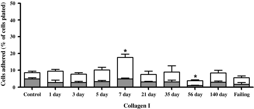
Fig. 5.
Left ventricular cardiomyocyte adhesion to type I collagen for various times postfistula and for a group with overt failure. Data presented as a percent of total cells plated. The shaded portion represents the residual adhesion following a preincubation with β1-integrin antibody. Means are ± SE. *P ≤ 0.05 vs. control.

