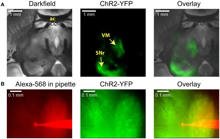
Figure 1.
Anatomical images illustrating a typical experiment. (A) Low-power images of a horizontal slice containing both the SNr and the VM. This level corresponds to Plate 144 in the Paxinos atlas (Franklin and Paxinos, 2008). AAV5 virus carrying YFP-tagged ChR2 under the control of the synapsin promoter was injected into the SNr, where it infected neurons and resulted in YFP fluorescence both in the surrounding tissue and in the VM thalamus. (B) Higher-power view shows the recording pipette and a VM neuron filled with the red dye Alexa-568 (left), the YFP fluorescence that marks ChR2 expression (middle), and the overlay (right). A glutamate puffer pipette can be seen on the left side of the middle image.

