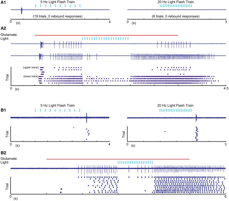
Figure 7.
Cell attached recordings with glutamatergic excitation and optical nigral input stimulation. (A1,B1) 5 Hz 10 ms and 20 Hz 5 ms light pulse trains result in LTS rebounds in one cell (B), but not another (A). Purple traces show recordings of extracellular spike currents and raster histograms below show spike times for repeated application of the same stimulus combination. (A2,B2) The same optical stimuli delivered during a 4 s 0.5 mM glutamate application result in pronounced spike pauses of glutamate-stimulated activity without a terminating LTS rebound burst in either cell. The strong initial rebound of cell (A) but not cell (B) with glutamate application suggests that this cell had a more hyperpolarized resting membrane potential than cell (B). A more depolarized baseline of cell (B) is also suggested by it showing LTS rebounds following IPSP trains as our intracellular recordings only resulted in such rebounds when the cell was considerably depolarized above ECl.

