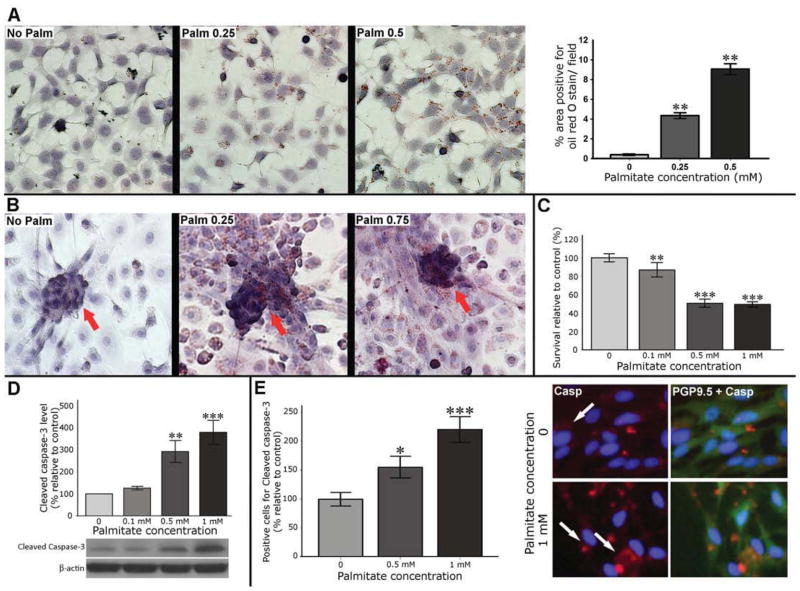
Figure 3. Palmitate is taken up by enteric neuronal cells and induces cell death.
(A) Representative microphotographs of neuronal cell line treated with palmita te and stained with oil red O. (B) Primary cell cultures of enteric neurons treated with palmitate with arrows indicate lipid deposits in the neuronal clusters. (C) MTS survival assay in enteric neurons after 24 hours of palmitate exposure. (D) Western blot analysis of Cleaved caspase-3 protein level in enteric neuronal cell lines. Graph depicts the average of five separate experiments. (E) IF staining of primary enteric neuronal cells. Arrows indicate cells positive for Cleaved caspase-3. Results presented as mean ± SEM, *P<0.05, ** P<0.01, ***P<0.001.

