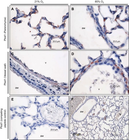
Fig. 2.
Localization of Plod1 expression in the parenchyma (A and B) and vessel walls (C and D) of the lungs of mouse pups at P7.5, after exposure to 21% O2 or 85% O2 from P0.5. The airways (aw) and vessels (v) are indicated. E: staining for Plod1 in a neonatal mouse lung at P7.5, after exposure to 85% O2 from P0.5, after preadsorption of the primary anti-Plod1 antibody with a competing peptide, to demonstrate specificity. F: low magnification staining for Plod1 in a neonatal mouse lung at P7.5 after exposure to 85% O2 from P0.5.

