
Fig. 6.
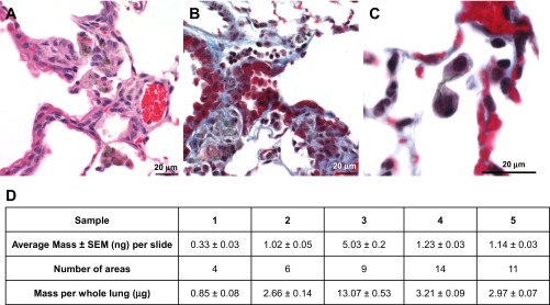
Photomicrographs of mouse lung 1 yr after inhalation of SWCNT (5 mg/m3, 5 h/day, 4 days, lung burden 5 μg). Photomicrographs presenting bronchointerstitial pneumonia (A) and peribronchiolar fibrosis with intralesional SWCNT compare the sites of peribronchiolar fibrosis with the location of the mildly dilated lymphatic (B, top of the photomicrograph) and cytokinesis failure (C) in SWCNT-exposed mice. D: amount of SWCNT retained in the lung tissue sections in mice after 1 yr postexposure as assessed by Raman spectroscopy.