
Fig. 9.
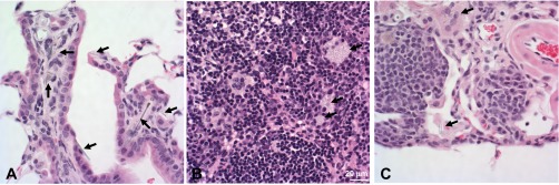
Photomicrographs 1 yr after pharyngeal aspiration with asbestos (120 μg/mouse). Photomicrographs demonstrating peribronchial inflammation, fibrosis, hyperplasia (A), granulomatous lymphadenitis in the paracortex of the tracheobronchial lymph node (B), pleuritis, and lymphangiectasia (C) after asbestos exposure.