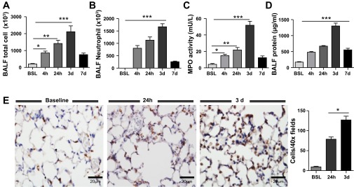
Fig. 2.

Time-dependent recruitment of neutrophils into the lung after intratracheal LPS. A and B: total immune cells and neutrophils in BAL fluid of control and LPS-injured lung. C: myeloperoxidase (MPO) activity in lung. D: total protein concentration in BAL fluid. E: neutrophil infiltration in the lung quantified by immunohistochemical staining for the granulocyte marker Gr-1. Gr-1+ cells (brown stain) peaked at 3 days after intratracheal LPS administration and declined thereafter. Morphometric analysis was performed on BSL, 24 h, and 3 days. Data are expressed as means ± SE of n = 6 animals in each group, *P < 0.05, **P < 0.01, and ***P < 0.001.
