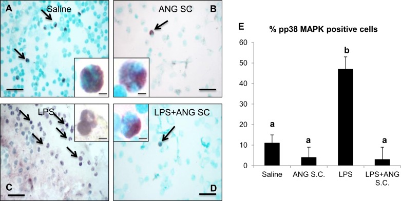
Fig. 11.
Phosphorylated p38 MAP kinase (pp38 MAPK) staining of peripheral blood neutrophil isolated from various mice groups, as described in materials and methods shows intense positive staining (marked by arrows) for LPS (C and inset) compared with saline (A and inset), subcutaneous angiostatin only (B and inset) and LPS+subcutaneous angiostatin (D and inset). Two-hundred peripheral blood neutrophils were counted from each group and plotted to represent the percentage of positive cells (E). a,bDifferent letters indicate statistical differences (P < 0.05) between the groups, while letters that match each other indicate no statistical difference. Scale bar = 10 μm for wide fields; 5 μm for insets.

