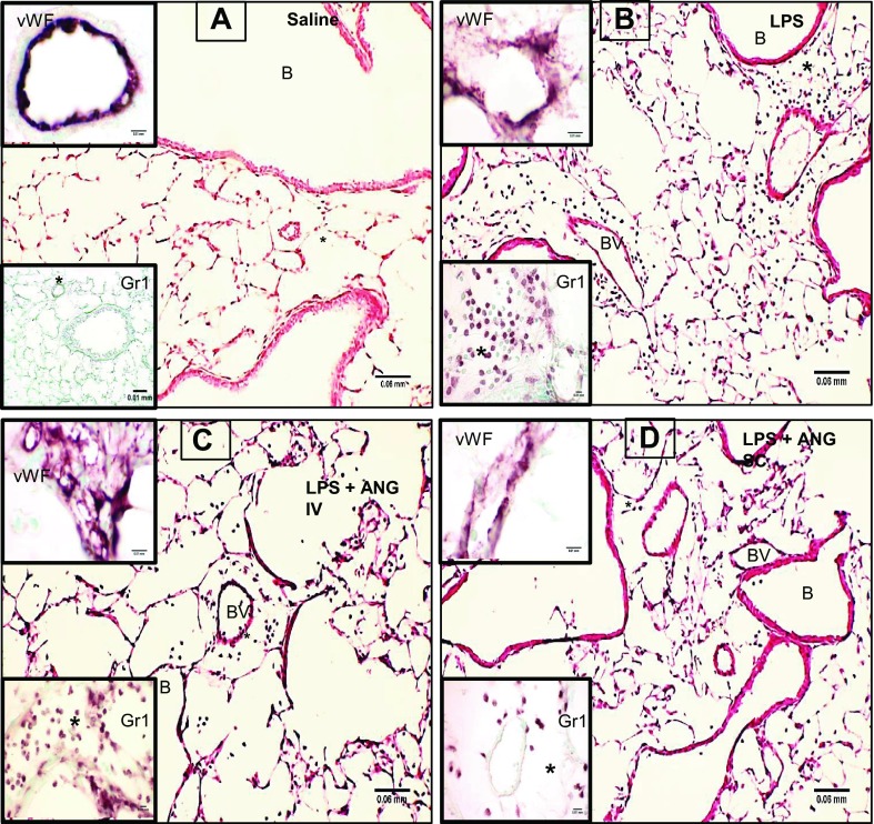
Fig. 3.
Hematoxylin-and-eosin (H&E)-stained lung sections from saline-treated control mouse show normal histology (A) and signs of inflammation in 9-h LPS-exposed mice (B). The inflammation at 9 h post-LPS treatment is much reduced in LPS-treated mice given subcutaneous angiostatin treatment (C) compared with those given angiostatin intravenously (D). The insets labeled vWF show expression of inflammatory marker von Willebrand Factor (vWF) to be more prominent in LPS+intravenous angiostatin compared with other groups. Gr1 immunohistochemistry insets show lack of neutrophils in perivascular compartment (*) in control mouse (A), while LPS-treated mouse lung showed robust accumulation of neutrophils in the perivascular compartment. The accumulation is nearly absent in lungs from mice given LPS followed by subcutaneous angiostatin (C) but not in the mice treated with angiostatin intravenously (D). BV, blood vessel; B, bronchiole.

