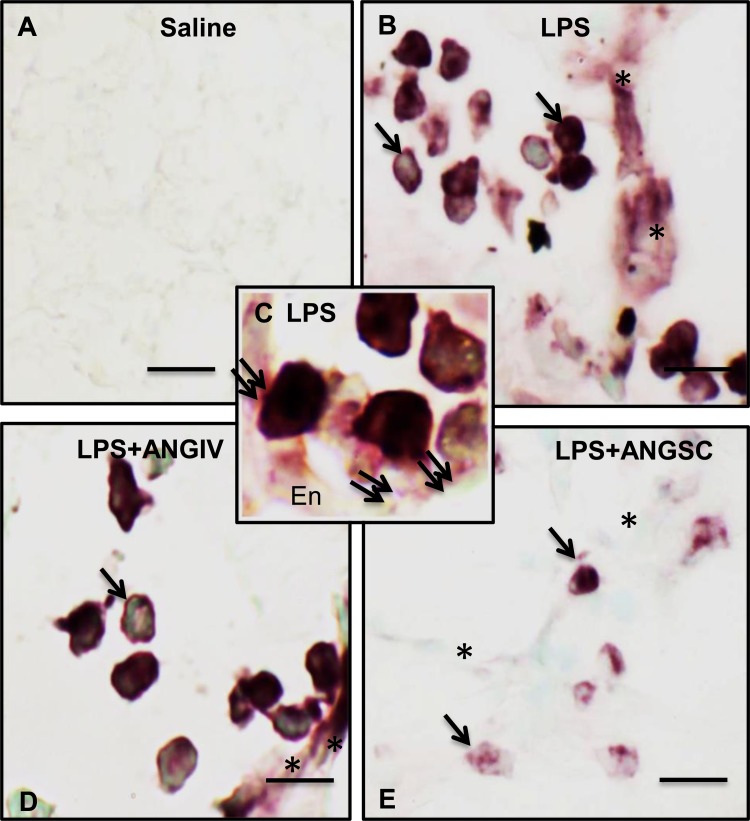
Fig. 6.
Phosphorylated p38 MAPK expression. The immunohistochemical protocol did not detect any staining for phosphorylated p38 MAPK in a section from control lung (A) but showed intense staining in many leukocytes (arrows) and septum (asterisks) in mice treated with LPS (B) or LPS+ANG IV (D). Higher magnification view (C) from an LPS-treated mouse lung shows leukocytes (double arrows) showing intense staining for phosphorylated p38 MAPK in contact with the endothelium (En). Lung section from a mouse treated with LPS+ANG SC (E) showed a few cells (single arrows) but not alveolar septum (asterisks) positive for phosphorylated p38 MAPK. Scale bar = 10 μm except bar in A = 50 μm.

