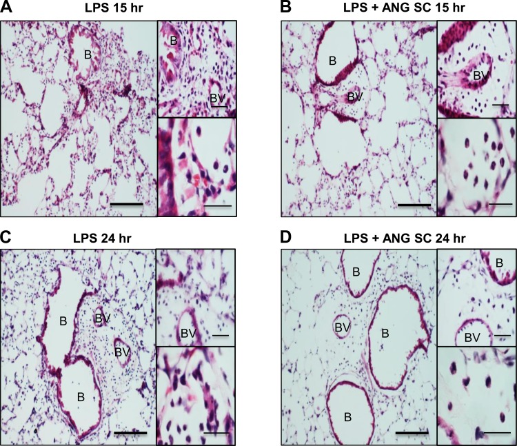
Fig. 9.
Hematoxylin-and-eosin-stained lung sections of mice at 15 h (A and insets) and 24 h (C and insets) show more inflammation compared with time-matched LPS+subcutaneous ANG-treated mice (B and D). The insets show reduced perivascular and alveolar neutrophils in LPS-challenged mice treated with subcutaneous angiostatin. Scale bar = 50 μm for wide fields; 20 μm for insets.

