
Fig. 2.
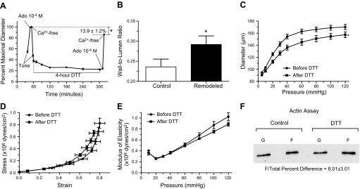
Prolonged exposure to DTT induces inward remodeling in isolated arterioles. A: arterioles were exposed to 200 μM DTT for 4 h. Before and after the 4-h exposure to DTT, arterioles were allowed to develop spontaneous tone and subsequently exposed to 10−4 M adenosine (Ado) and then to calcium-free solution. Data are means ± SE of the maximal passive diameter obtained during the first exposure to calcium-free conditions. After the second exposure to calcium-free conditions, maximal passive diameter was significantly reduced (*P ≤ 0.05; n = 5). B: wall/lumen ratios of arterioles in calcium-free conditions before (control) and after a prolonged (4-h) exposure to DTT (n = 5). *P ≤ 0.05 vs. control. C: passive pressure-diameter curves of arterioles obtained before (before DTT, n = 5) and after (after DTT, n = 5) exposure (4 h) to 200 μM DTT. *P ≤ 0.05 vs. before DTT. D: passive strain-stress relationships of isolated arterioles before (before DTT, n = 5) and after (after DTT, n = 5) exposure (4 h) to 200 μM DTT. E: incremental modulus of elasticity vs. pressure in isolated arterioles under passive conditions before (before DTT, n = 5) and after (after DTT, n = 5) exposure (4 h) to 200 μM DTT. F: representative immunoblot of the F- and G-actin portions of the cytoskeleton from arterioles treated with vehicle control or DTT (200 μM) for 4 h. Exposure to DTT increased (P ≤ 0.05) the F-actin/total actin by 8.01 ± 3.01% vs. controls (n = 6 for each treatment).