Fig. 3.

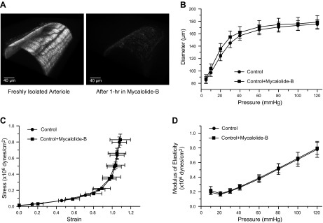
Actin cytoskeletal disruption does not affect the passive diameter or elastic characteristics of freshly isolated arterioles. A: 3-dimensional confocal images of isolated arterioles exposed to vehicle control (left) or 2 μM mycalolide-B (right) and subsequently stained with phalloidin-Alexa 546 to visualize the actin cytoskeleton. B: pressure-diameter curves of freshly isolated arterioles before (control, n = 7) and after (control + mycalolide-B, n = 7) exposure (1 h) to 2 μM mycalolide-B. C: strain-stress relationships of freshly isolated arterioles before (control, n = 7) and after (control + mycalolide-B, n = 7) exposure (1 h) to 2 μM mycalolide-B. D: incremental modulus of elasticity vs. pressure in freshly isolated arterioles before (control, n = 7) and after (control + mycalolide-B, n = 7) exposure (1 h) to 2 μM mycalolide-B.
