Fig. 1.
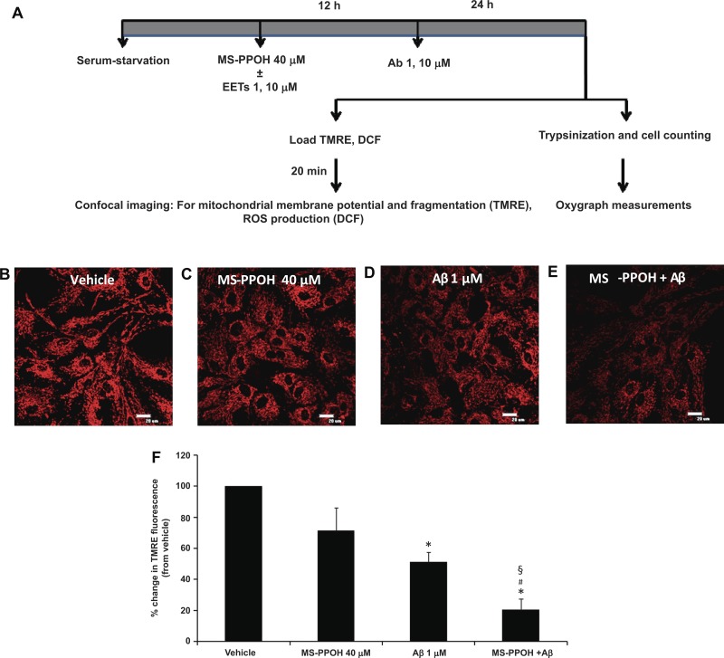
Preincubation with MS-PPOH aggravates amyloid-β (Aβ, Ab)-induced mitochondrial membrane depolarization. A schematic diagram depicting experimental procedure (A). EETs, epoxyeicosatrienoic acids; DCF, 5 (and 6)-chloromethyl-2′,7′-dichlorodihydrofluorescein diacetate acetyl ester; ROS, reactive oxygen species. For mitochondrial membrane potential measurements astrocytes were loaded with tetramethylrhodamine ethyl ester perchlorate (EET; which localizes to the inner mitochondrial membrane) after being treated with vehicle (B), MS-PPOH (C), Aβ (D), and MS-PPOH + Aβ (E) (×60 magnification; scale bar = 20 μm). The relative change in fluorescence intensity is expressed as percent change from vehicle set at 100%. Aβ (1 μM) causes a decrease in mitochondrial membrane potential (seen as a decrease in EET fluorescence) compared with vehicle, which is further decreased by incubation with MS-PPOH (F). *P < 0.001 vs. vehicle; #P < 0.05 vs. Aβ; §P < 0.001 vs. MS-PPOH; n = 5 to 6.

