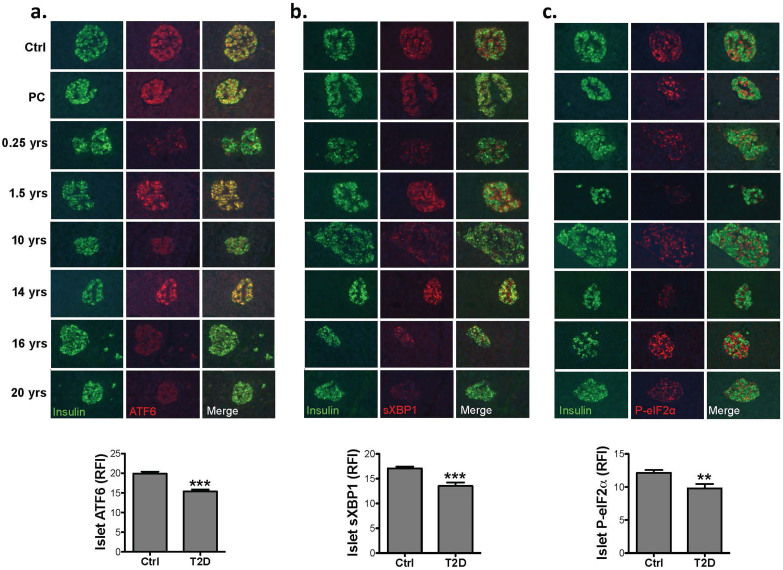
Figure 3. Expression of UPR mediators in the islets of type 2 diabetic humans.
(a). Pancreas sections from non-diabetic and diabetic patients were obtained from nPOD and co-stained with anti-ATF6α (red), and anti-insulin (green) antibodies (upper panel) and 10–20 islets per sample were quantified by MATLAB® (lower panel). (b). Pancreas sections from non-diabetic and diabetic subjects co-stained with anti-sXBP1 (red) and anti-insulin (green) antibodies (upper panel) and 10–20 islets per sample were quantified by MATLAB® (lower panel). (c). Pancreas sections from non-diabetic and diabetic subjects co-stained with anti-P-eIF2α (red) and anti-insulin (green) antibodies (upper panel) and 10–20 islets per sample per time point were quantified by MATLAB® (lower panel). All data are presented as mean ± SEM, with statistical analysis performed by one-way ANOVA (***p < 0.001, **p < 0.005, *p < 0.05).

