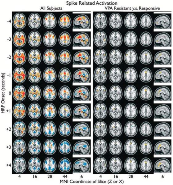
Figure 1.

Spike-related activation (red) and deactivation (blue), controlling for number of spikes, are overlayed on the standard MNI anatomic brain in radiologic orientation. Spike times obtained from a simultaneous EEG recording were convolved with a canonical gamma variate hemodynamic response function (HRF)with its peak 4.7 s post onset using AFNI. Activation maps were produced with HRF onset aligied with spikes (0 s), before spikes (−1,−2, … s), and after spikes (+1,+2, … s). Group responses from all subjects (left) and a contrast between vdproate (VPA)-resistant and VPA-responsive subjects(right) were obtained. Significant (a < 0.05) clusters of >36 suprathreshold voxels (left: two-sided t > 2.3, right: one-sided t > 2) are shown.
Epilepsia © ILAE
