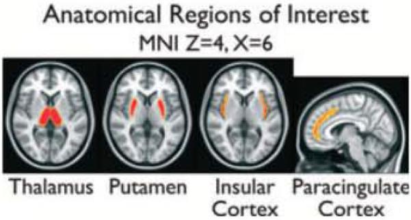
Figure 2.

Slices from the anatomic regions of interest (ROIs) used in Figs 3 and 4 are overlayed on the standard MNI anatomic brain In radiologic orientation. Regions were obtained from the Harvard-Oxford cortical and subcortical probabilistic atlases distributed with FSL Voxels with probability >50% of residing within the anatomic region are shown and were used as a mask to compute the ROI time courses in Figures 3 and 4.
Epilepsia © ILAE
