
Fig. 2.
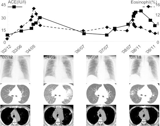
Clinical course from December 2002 to October 2010. Chest Xp and CT showed consolidation in left lung field in 2004; additionally, serum ACE activity and percentage of peripheral eosinophil were elevated. In 2008, ground-glass opacities were evident in the right lung filed, and serum ACE activity and the percentage of peripheral eosinophil were again elevated, but spontaneously remitted.