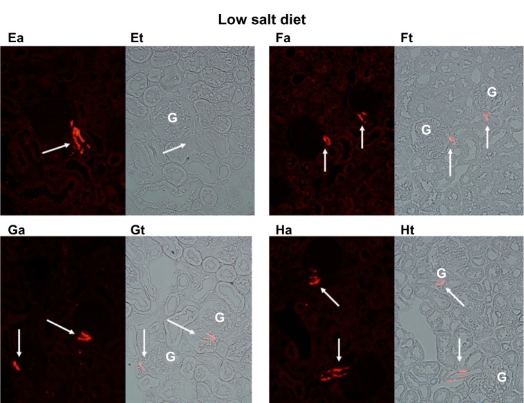
Figure 2.
Immunofluorescence of rat renal cortical sections fixed in situ on the left (a) and a paired light-transmission micrograph (t) of the same field on the right. The fluorescence images show (in red) renin extending up the afferent arteriole (indicated by white arrows) from the glomerulus (G) shown at 40×. This figure depicts four examples (E–H) from kidneys of rats maintained on a sodium-restricted diet over 12 days, in which renin recruitment is clearly seen extending from the juxtaglomerular apparatus (JGA) up the afferent arteriole away from the JGA.

