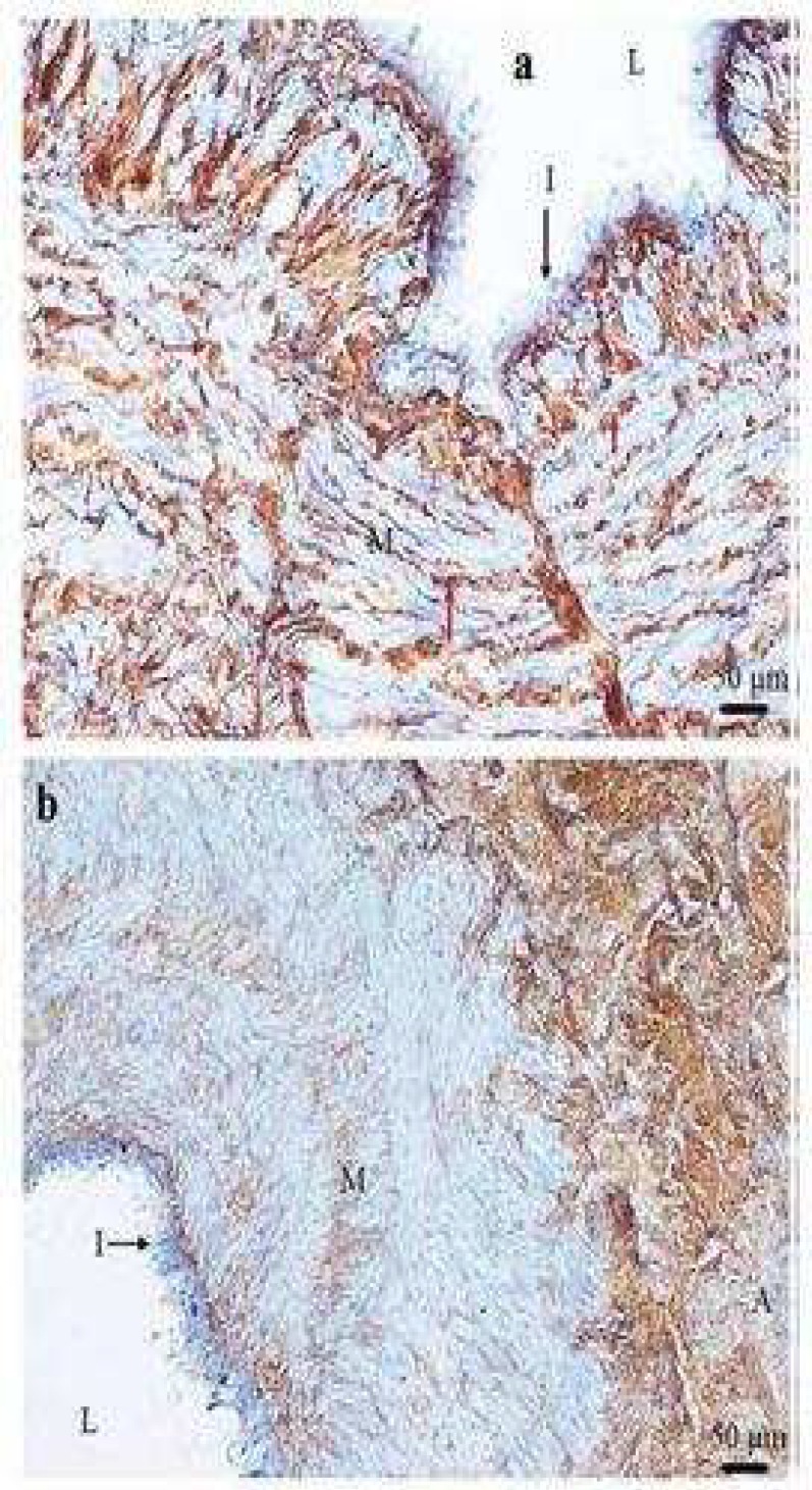
Fig 5.
Type III collagen expression in control saphenous vein (a) and varicose vein (b). Typical transverse section of control saphenous vein (a) and varicose veins (b) stained with type III collagen antibody showing strong and weak positive staining (brown) respectively. Strong positive staining was seen in the media (M) and scattered between smooth muscle cells (red arrows). The adventitia (A) shows strong positive staining of type III collagen. I: intima; L: lumen of the vein (×200) (×100

