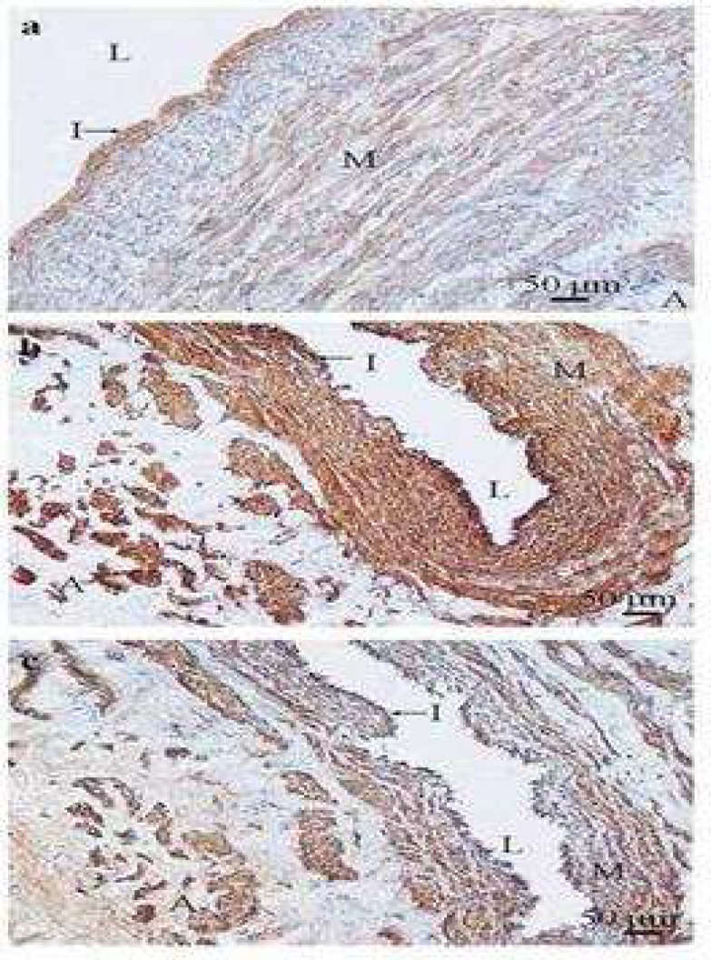
Fig 6.
Type IV collagen expression in control saphenous vein and Type IV collagen expression compared with α- smooth muscle actin expression in varicose vein. Typical transverse section of control saphenous vein (a) stained (brown) with type IV collagen antibody showing strong positive staining in the intima (I). The media (M) shows positive staining for type IV collagen in the smooth muscle cell basement membranes. Typical transverse section of the same varicose vein stained with type IV collagen (b) and α -smooth muscle actin (c) antibodies. The positive staining (brown) of type IV collagen was found in the intima (I) which indicated the type IV collagen components of basement membrane (arrow). Type IV collagen components of basement membrane were also detected smooth muscle cell basement membranes in the media (M) and the adventitia (A). The immunostaining distribution pattern of both type IV collagen (b) and α -smooth muscle actin (c) antibodies staining indicated the role of type IV collagen as a component of basement membrane in the media and the adventitia which surrounds smooth muscle cells. L= lumen of the vein (×100

