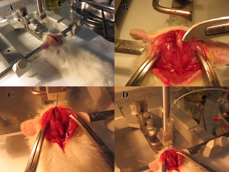
Figure 1.
A: Rat was placed in a stereotaxic head holder. The head was flexed downward at approximately 90◦. A midline scalp incision was made and the cervico-spinal muscle was reflected. B: The atlanto-ocipital membrane exposed. C: The atlanto-occipital membrane punctured by the fire-polished 1 ml syringe connected to the 27G dental needle. D: Using a special stereotaxic guide to hold the syringe

