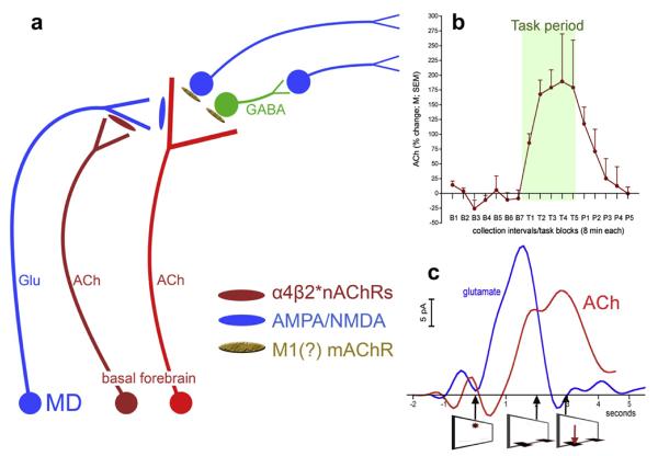
Fig. 1.
Schematic illustration of the main elements of prefrontal (PFC) circuitry mediating the detection of cues and of the tonic (dark red cholinergic neuron) and phasic (bright red cholinergic neurons) components of cholinergic neurotransmission (a). In the cortex, cholinergic transients are generated in part by the stimulation of ionotropic glutamate receptors and by glutamate released from mediodorsal thalamic (MD) afferents (Parikh et al., 2008, 2010). Mediodorsal inputs and thus glutamatergic transients are modulated by tonic (changes occurring on the scale of minutes) cholinergic stimulation of α4β2* nicotinic acetylcholine receptors (nAChRs). This allows levels of motivation and the subjects's readiness for utilizing external cues to influence the probability and efficacy of cue detection. An example of such tonic cholinergic activity measured by using microdialysis is shown in b, illustrating tonic prefrontal ACh before, during, and after four blocks of 8-min trials of sustained attention performance (data adopted from Paolone et al., 2010). In the PFC, glutamatergic and cholinergic transients interact to recruit efferent circuitry for the generation of the behavioral response. Figure c shows prefrontal glutamatergic and cholinergic transients recorded during cued trials yielding hits (the onset of the cue and the times of lever extension and correct lever presses are indicated below the abscissa). Note that the timing of detection-indicating behavior is related to the timing of the initial rise in transient cholinergic activity, as opposed to transient peak time (Howe and Sarter, 2010; Parikh et al., 2007).

