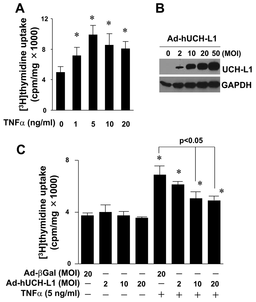
Fig. 1.
Effect of UCH-L1 over-expression on TNFα-induced VSMC proliferation. (A) TNFα-induced proliferation of rat aortic smooth muscle cells (RASMCs). Cell proliferation was assessed by measuring [3H]thymidine update as described in “Methods”. * p<0.05 vs TNFα (−), n=4. (B) Adenoviral over-expression of UCH-L1 in RASMCs. Results are representative of three independent Western blot analysis of UCH-L1 in RASMCs infected with or without adenovirus of UCH-L1. (C) Effect of over-expression of UCH-L1 on TNFα-induced RASMC proliferation. Cells infected with Ad-UCH-L1 or Ad-βGal were stimulated with or without TNFα (5 ng/ml) as indicated for 40 hours. * p<0.05 vs TNFα (−), n=4.

