Figure 2.
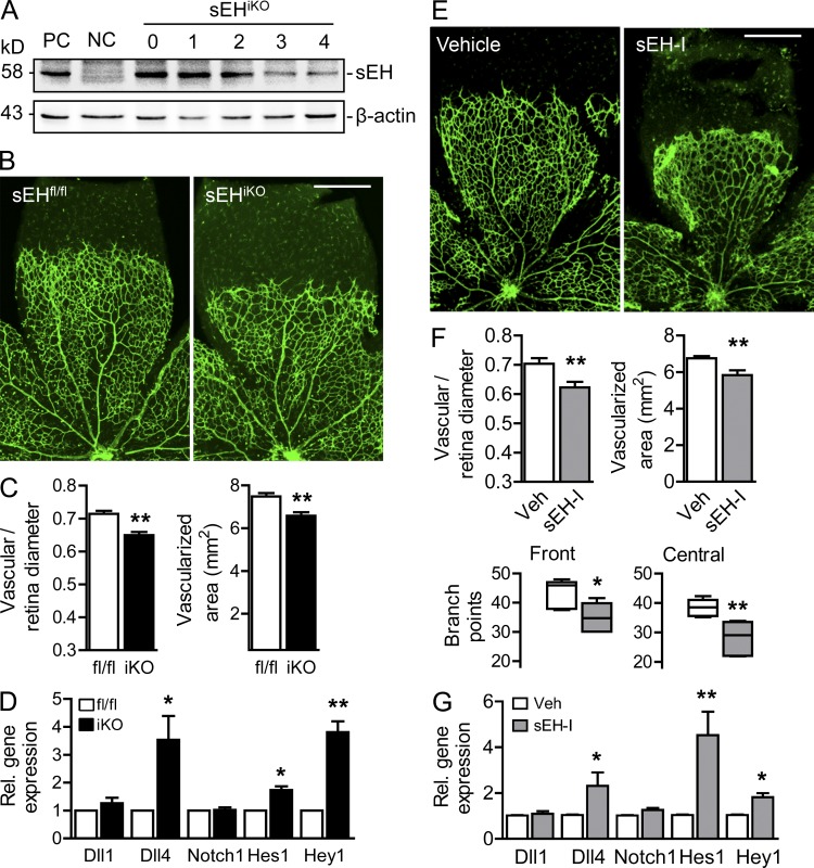
Consequences of the postnatal down-regulation and inhibition of the sEH on retinal angiogenesis. (A) sEHiKO mice were treated with tamoxifen from P1 to P4 and sEH expression was determined by Western blotting. Similar observations were obtained in 5 additional experiments. Retinas from wild-type and sEH−/− mice were used as positive (PC) and negative (NC) controls, respectively. (B) Isolectin B4 staining in retinal whole mounts from tamoxifen-treated sEHfl/fl and sEHiKO mice on P5. Bar, 500 µm. (C) Quantification of vascularization. n = 5 total animals per group, from 3 different experiments. (D) Expression of genes involved in Notch signaling was assessed in P5 retinas by RT-qPCR. Each sample was a pool of 3 retinas, and the analysis was performed on 4 independent samples per genotype. (E) Wild-type mice were treated with vehicle (Veh) or sEH inhibitor (sEH-I) for 4 d and Isolectin B4 staining was analyzed by confocal microscopy. Bar, 500 µm. (F) Quantification of vascularization based in data in E. n = 5–9 total animals per group from 4 different experiments. (G) 5-d-old wild-type mice were treated with vehicle or sEH inhibitor for 4 d, and Notch pathway gene expression was assessed by RT-qPCR. Each sample was a pool of 3 retinas, and the analysis was performed on 6 independent samples per genotype. Error bars represent SEM. *, P < 0.05; **, P < 0.01 versus sEHfl/fl mice or vehicle.

