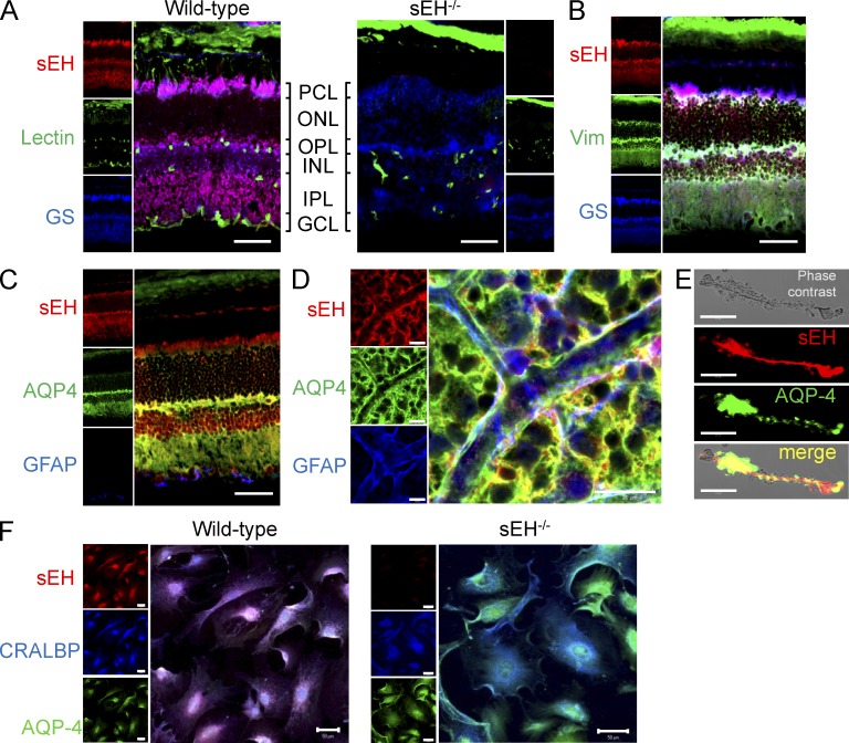
Figure 3.
Localization of sEH in the retina. (A) sEH (red), Isolectin B4 (green), and GS (blue) levels in retinal cryosections from wild-type and sEH−/− mice were assessed by confocal microscopy. GCL, ganglion cell layer; IPL, inner plexiform layer; INL, inner nuclear layer; OPL, outer plexiform layer; ONL, outer nuclear layer; PCL, retinal pigment cell layer. Bars, 50 µm. (B and C) sEH and vimentin (Vim) and GS (B) or AQP-4 and GFAP (C) expression in wild-type retinas was assessed by confocal microscopy. Bars, 50 µm. Similar observations were made in 4 additional experiments. (D) sEH, AQP-4, and GFAP expression in whole mounts of wild-type retinas assessed by confocal microscopy. Bars, 20 µm. Similar observations were made in retinas from 4 additional experiments. (E) sEH and AQP-4 expression in freshly isolated Müller cells from a wild-type retina. Bars, 20 µm. Similar observations were obtained in 4 independent experiments each using cells isolated from different litters. (F) Expression of sEH, CRALBP, and AQP-4 in primary cultures of Müller cells isolated from wild-type and sEH−/− retinas. Bars, 50 µm. Similar observations were obtained in 4 independent experiments each using cells isolated from different litters.

