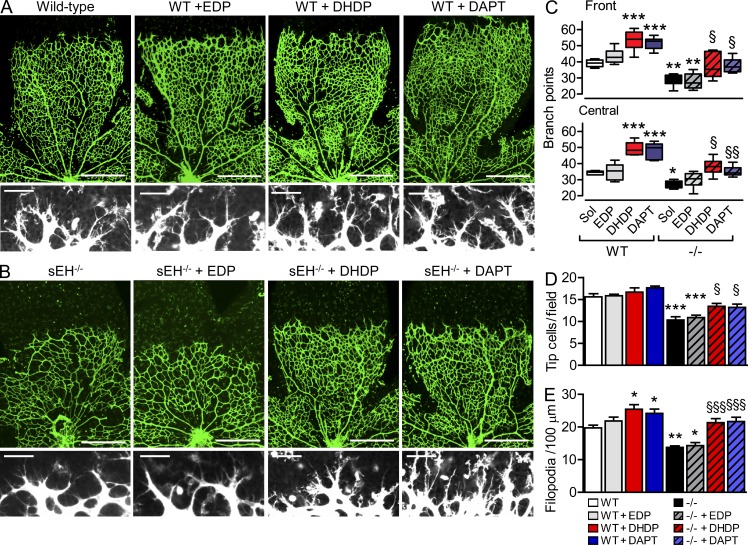
Figure 9.
19,20-DHDP promotes retinal angiogenesis in vivo. (A and B) Isolectin B4–stained retinal vasculature (top; bars, 500 µm) and tip cells at the angiogenic front (bottom; bars, 50 µm) of P5 retinas was assessed by confocal microscopy of whole mounts from wild-type (WT) mice (A) or sEH−/− (−/−) mice (B) 5 h after intravitreal injection of 0.5 µl solvent, 100 µmol/liter 19,20-EDP, 100 µmol/liter 19,20-DHDP, or 200 µmol/liter DAPT. (C–E) The graphs summarize vessel branch points (C), tip cell (D), and filopodia (E) numbers; n = 1–2 mice/group, and experiments were independently performed 3 times. Error bars represent SEM. *, P < 0.05; **, P < 0.01; ***, P < 0.001 versus wild-type. §, P < 0.05; §§, P < 0.01; §§§, P < 0.001 versus sEH−/− and solvent.

