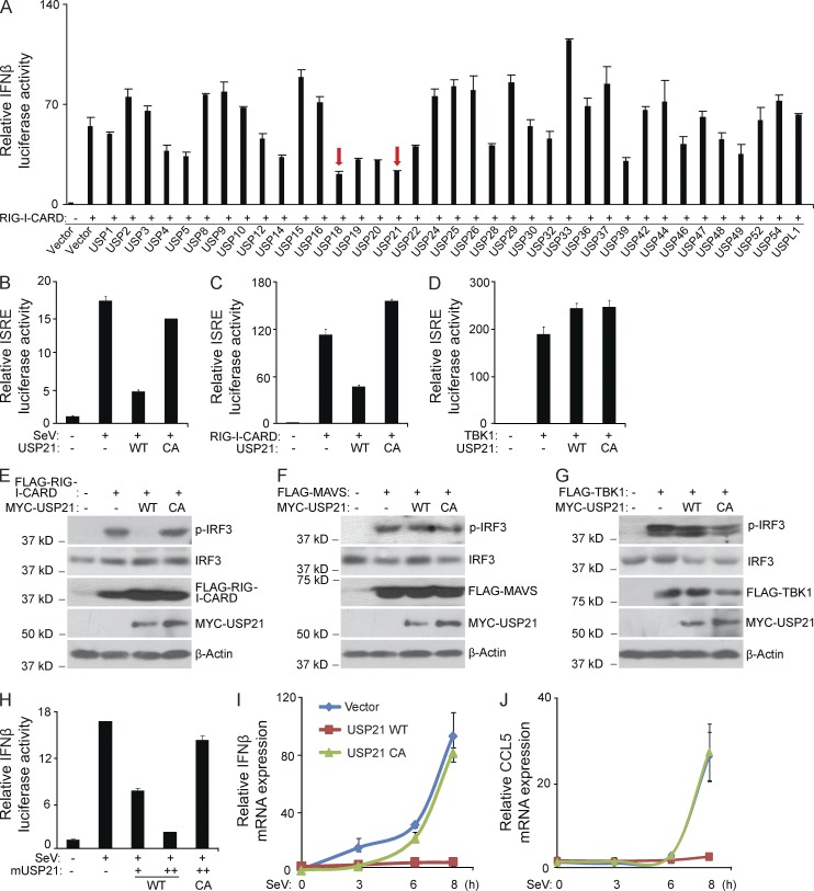
Figure 1.
USP21 inhibits RIG-I-CARD–induced IRF3 activation and IFN-β signaling. (A) RIG-I-CARD was co-transfected with indicated USPs or control vectors along with IFN-β luciferase reporter into HEK293T cells for 36 h. IFN-β luciferase activity was measured and normalized with the Renilla activity. (B) HEK293T cells were transfected with USP21 WT or C221A (CA) mutant along with ISRE luciferase reporter vectors for 24 h, and then infected with SeV or left uninfected for 12 h before ISRE luciferase was measured. (C and D) RIG-I-CARD or TBK1 were co-transfected with USP21 WT or CA mutant along with ISRE luciferase reporter vectors into HEK293T cells for 36 h before ISRE luciferase activity was measured. (E–G) FLAG-RIG-I-CARD, FLAG-MAVS, or FLAG-TBK1 were co-transfected with empty vector or expression vector encoding MYC-USP21 WT or CA mutant into HEK293T cells for 36 h. Cells lysates were immunoblotted with the indicated antibodies. (H) HEK293T cells were transfected with mouse orthologous USP21 WT or CA mutant along with IFN-β luciferase vectors for 24 h, and then infected with SeV or left untreated for 12 h before luciferase was measured. (I and J) HEK293T cells were transfected with USP21 WT or CA mutant for 36 h, and then infected with SeV at the indicated time points. The mRNA level of IFN-β and CCL5 was measured by real-time RT-PCR. Error bars indicate ±SD in duplicate experiments. Data from A is a screening data from one experiment. Data are representative of two (H–J) or at least three (B–G) independent experiments.

