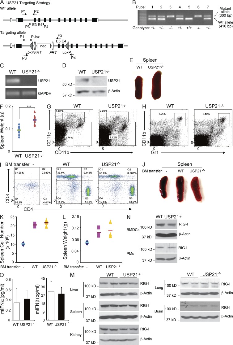
Figure 5.
Generation of USP21 knockout mice. (A) Generation of USP21 conditional knockout mice by two-loxP, two-frt strategy. (B) PCR genotyping of WT and USP21−/− littermates generated from USP21+/− mice intercrossing. (C) Total RNA was extracted from MEFs and mRNA level of USP21 was examined by RT-PCR. (D) Cell lysates from WT and USP21−/− MEFs were immunoblotted by anti-USP21 antibodies. (E) Representative images of spleens isolated from 9-wk WT and USP21−/− mice. (F) Quantitative analysis of the weight of spleens isolated from 9-wk-old WT (n = 10) and USP21−/− (n = 10) mice. (G) Macrophage and dendritic cell populations in splenocytes from USP21−/− and WT mice were analyzed by FACS using anti-CD11b and anti-CD11c antibodies. (H) Neutrophil populations in splenocytes from USP21−/− and WT mice were analyzed by FACS using anti-Gr1 and anti-CD11b antibodies. (I) SCID mice were transferred with WT or USP21−/− BM cells for 10 wk. T cell populations in splenocytes were analyzed by FACS using anti–mouse CD3, CD4, and CD8 antibodies. (J) Representative images of spleens from naive and BM-transferred SCID mice. (K) Spleens from naive and BM transferred SCID mice were homogenized. Total splenocytes were quantitatively analyzed. (L) Quantitative analysis of the weight of spleens isolated from naive and BM-transferred SCID mice. (M) Fresh BM cells were cultured in the medium with IL-4 and GM-CSF cytokines for 7 d. Cell lysates from BMDCs or PMs were immunoblotted by anti–RIG-I antibodies. (N) Blood was collected from 9-wk-old WT or USP21−/− mice, and IFN level in sera was measured by ELISA kit. (O) Cells from different organs were lysed and immunoblotted by anti–RIG-I antibodies. Error bars indicate ±SD in duplicate experiments. ***, P < 0.001 (two-tailed paired Student’s t test). Data are representative of two (E–O) or at least three (B–D) independent experiments.

