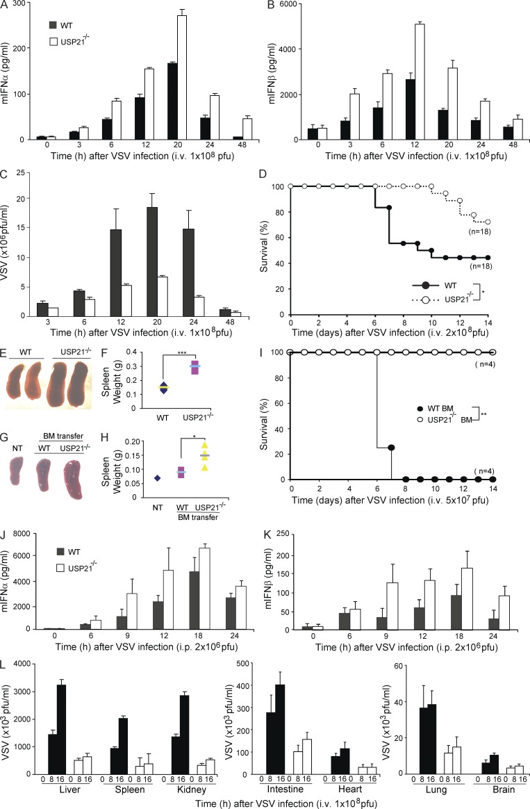
Figure 9.
Knockout of USP21 expression enhances antiviral response in vivo. (A and B) WT and USP21−/− mice were injected with VSV (1 × 10⁸ pfu) via tail vein for the indicated time period. Amounts of IFN-α and IFN-β in sera were measured by ELISA. (C) WT and USP21−/− mice were infected with VSV (1 × 10⁸ pfu) via tail vein injection. Sera collected at the indicated time points were used for measurement of viral titers by plaque assays. (D) WT (n = 18) and USP21−/− (n = 18) mice were infected with VSV (2 × 10⁸ pfu) via tail vein injection and the survival of the mice was monitored for 2 wk. (E) Representative images of spleens isolated from WT and USP21−/− mice 2 wk after intravenous VSV (2 × 10⁸ pfu) infection. (F) Quantitative analysis of the weight of spleens isolated from 9-wk-old WT (n = 5) and USP21−/− (n = 5) mice 2 wk after intravenous VSV (2 × 10⁸ pfu) infection. (G and H) SCID mice were transferred with WT and USP21−/− BM cells and infected with VSV (1 × 10⁷ pfu) for 2 wk. Representative spleens (G) were shown and spleen weight (H) was measured. (I) SCID mice transferred with WT and USP21−/− BM cells were infected with VSV (5 × 10⁷ pfu) via tail vein injection and the survival of the mice was monitored for 2 wk. (J and K) WT and USP21−/− mice were injected intraperitoneally with VSV (2 × 10⁶ pfu) for the indicated time courses. Amount of IFN-α (J) and IFN-β (K) in sera was measured by ELISA. (L) WT and USP21−/− mice were injected with VSV (1 × 10⁸ pfu) through tail vein for the indicated time points. Mice were sacrificed and viral titers in organs were determined by the plaque assay. Error bars indicate ±SD in duplicate experiments. *, P < 0.05; **, P < 0.01; ***, P < 0.001 (two-tailed paired Student’s t test or Kaplan Meier survival analysis). Data are representative of two (A–C and E–L) or at least three (D) independent experiments.

