
Fig. 2.
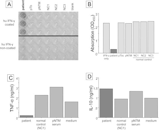
(A) Antigen capture assays of immunoglobulins against IFN-γ in our patient's serum (left lane), a pulmonary tuberculosis (pTb) patient's serum (2nd lane from the left), a pulmonary NTM (pNTM) patient's serum (3rd lane from the left), and normal control (NC) sera (4th–6th lanes from the left). In each lane, the upper 3 wells were coated with recombinant human (rh) IFN-γ and the lower 3 wells were not. The bound immunoglobulins were detected using horseradish peroxidase conjugated anti-human Fab specific antibody. (B) Inhibition binding activity of anti-IFN-γ autoantibody was as described in the “Case report” section. All samples include 100 pg of IFN-γ and sera from patients, patients with pTb and pNTM, and normal controls except the 1st lane from the left. The sample of the 1st lane contained 100 pg of IFN-γ only with PBS. All added sera were the same as those used for the antigen capture assays. The vertical axis is absorption of IFN-γ, as shown by optical density at 450 nm (OD450). (C) and (D) Productions of TNF-α and IL-10 by peripheral blood mononuclear cells (PBMNCs) were measured using sera from a normal individual, as described in the “Case report” section. In total, 1.0 × 10⁶ PBMNCs in a total of 2 mL of complete medium (RPMI1640 and 10%/v fetal calf serum) were incubated with LPS (200 ng/ml) and IFN-γ (1 ng/ml) in the presence of 100 μl of our present patient's serum, as well as sera from other previous pNTM patients and normal controls. All added sera were the same as those used for the antigen capture assays.