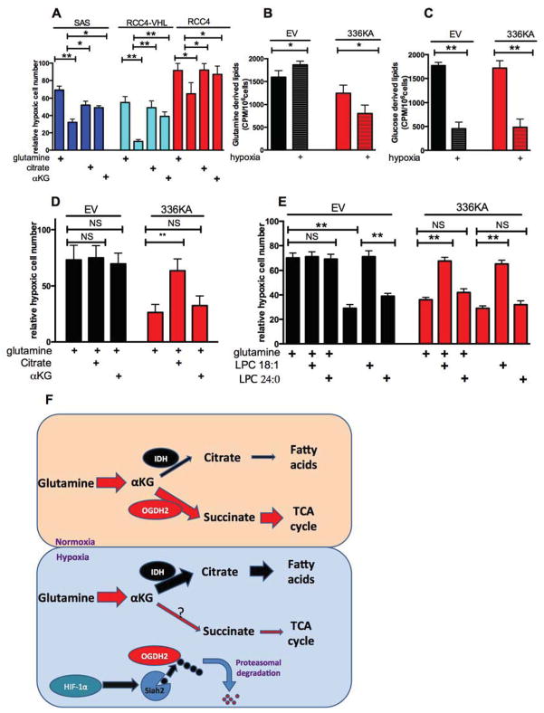
Figure 4. OGDH 336KA inhibits cell growth under hypoxia by reducing glutamine derived lipid production, see also figure S4.
Panel A: Relative hypoxic proliferation of SAS, RCC4 and RCC4VHL cells grown for 72 h in the indicated medias, presented as a percentage of normoxic growth. Hypoxic growth requires either glutamine (2mM), or glutamine derivatives αKG (2mM) or citrate (2mM).
Panel B Hexane soluble lipids derived from a 1h pulse of 0.5 μCi 14C-glutamine in SAS cells expressing either empty vector or OGDH2-336KA, grown for 16h in normoxia or hypoxia.
Panel C. Hexane soluble lipids derived from a 1h pulse of 0.5 μCi 14C-glucose in SAS cells as described in B.
Panel D. Relative hypoxic proliferation of SAS cells expressing empty vector or OGDH2-336KA after 72 hours in basal glutamine-containing media and 10% charcoal-stripped serum supplemented either 2mM citrate or 2mM αKG. Note WT cells have maximal hypoxic proliferation with glutamine, but 336KA OGDH2 expressing cells require citrate.
Panel E Relative hypoxic proliferation of SAS cells described in D, in basal media with 10% charcoal-stripped serum. Supplementation with absorbable lysophospholipid rescues the glutamine dependence of the WT OGDH2 cells, and the citrate dependence of the 336KA OGDH2 expressing cells. Data from panels A–E represents mean +/− S.D.
Panel F. A model illustrating how hypoxic degradation of OGDH2 shifts the fate of αKG from energy production to the production of lipids.

