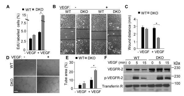
Figure 2. Epsin deficiency promotes VEGF-dependent in vitro angiogenesis.
A, Quantification of EdU incorporation into WT or DKO MECs after labeling in the absence or presence of VEGF-A (50 ng/mL) for 24 h. B, WT or DKO MEC monolayers were subjected to a scratch assay in the absence or presence of VEGF-A (50 ng/ml) for 12 h. C, Wound distance in B at 12 h was quantified using NIH ImageJ software. D, WT or DKO MECs were subjected to a tube formation assay by culturing on matrigel for 16 h in the absence or presence of VEGF-A (50 ng/ml). E, Tube formation in D at 16 h was quantified as in C. F, Plasma membrane fractions from WT or DKO MECs stimulated with 50 ng/mL VEGF-A for 0, 5 and 15 min were immunoblotted using the indicated antibodies. A, C and E, error bars indicate the mean ± s.e.m. n > 5, *p < 0.05. Scale bars: B and D, 50 μm.

