Figure 4.
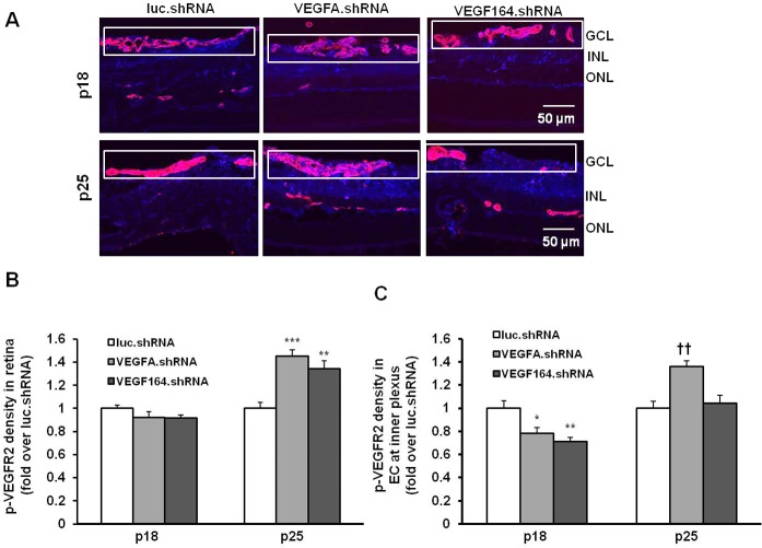
Analysis of VEGFR2 activation in pups treated with subretinal injections of lentivector-driven shRNAs in the rat ROP model. (A) Immunohistochemistry of p-VEGFR2 in retinal cryosections. (B) Semiquantification of p-VEGFR2 (blue) in total retina (***P < 0.001 versus luc.shRNA at P25). (C) Colabeling of p-VEGFR2 (blue) in lectin (red)-labeled ECs in the primary plexus (depicted within boxes; *P < 0.05, **P < 0.01 versus luc.shRNA at P18; ††P < 0.01 versus luc.shRNA at P25) from P18 and P25 pups treated with luc.shRNA, VEGFA.shRNA, and VEGF164.shRNA.

