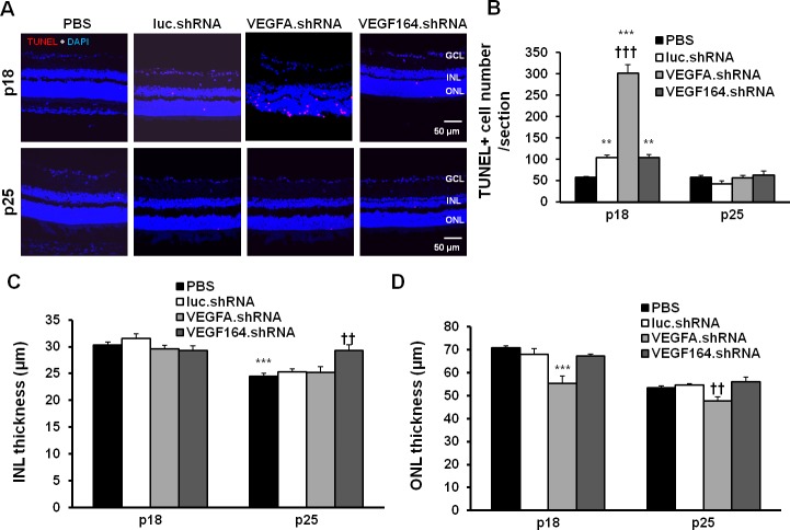
Figure 5.
Analysis of retinal apoptosis and retinal morphological changes in the pups treated with lentivector-driven shRNAs in the rat ROP model. Images of TUNEL staining (A) and number of TUNEL (red) positive cells (B) in retinal DAPI (blue) stained cryosections from P18 and P25 pups treated with luc.shRNA, VEGFA.shRNA, and VEGF164.shRNA (**P < 0.01, ***P < 0.001 versus PBS at P18; †††P < 0.001 versus luc.shRNA at P18). Quantification of the thickness of the INL (C) (***P < 0.001 versus PBS at P18; ††P < 0.01 versus luc.shRNA at P25) and the ONL (D) (***P < 0.001 versus luc.shRNA at P18; ††P < 0.001 versus luc.shRNA at P25) in DAPI-stained retinal cryosections from P18 and P25 OIR pups treated with luciferase.shRNA, VEGFA.shRNA, and VEGF164.shRNA.

