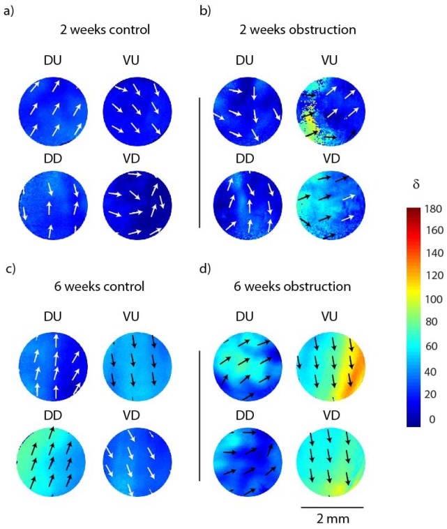
Fig. 3.
Representative regional retardances (anisotropy) obtained from polarized light imaging and Mueller matrix decomposition of a) a 2-week control rat bladder, b) a 2-week obstructed bladder, c) a 6-week control bladder, d) a 6-week obstructed bladder. DU = dorsal urethral, DD = dorsal dome, VU = ventral urethral and VD = ventral dome (anatomically depicted in Fig. 1). Color bar shows retardance values in degree and the arrows show the orientation of the retardance (optical axis).

