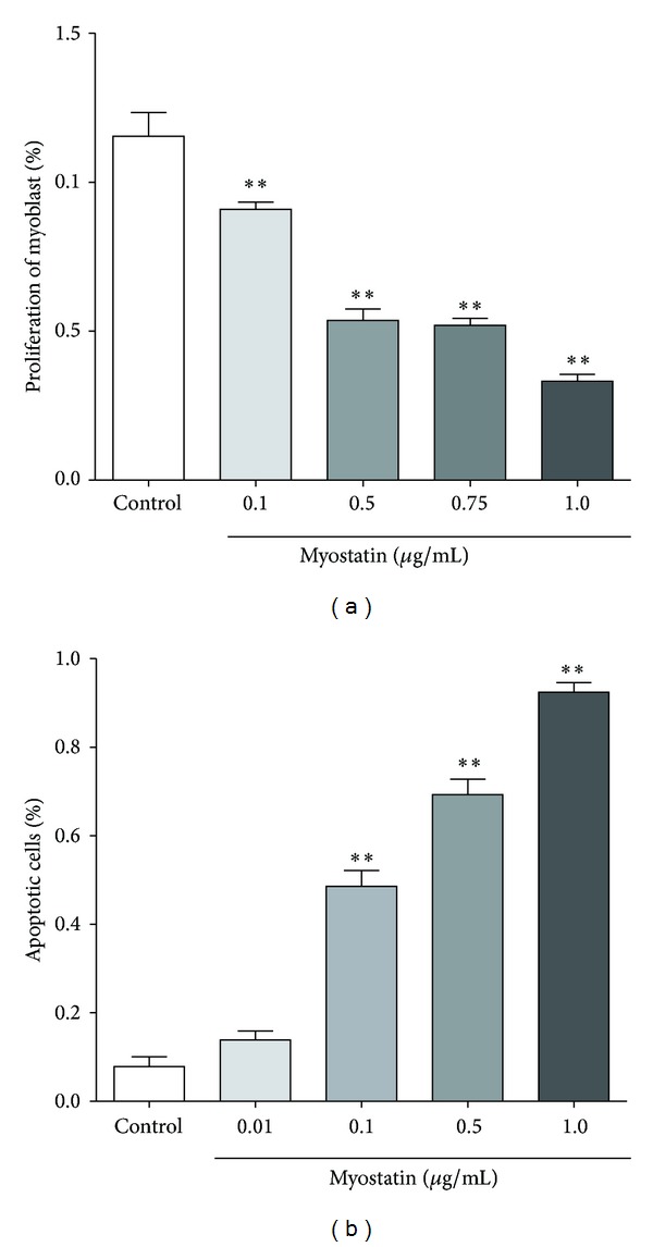
Figure 4.

(a) Proliferation of murine myoblasts is shown as percentage after exposure to different myostatin concentrations for 24 h. Increasing concentrations of myostatin were associated with decreased proliferation rate of murine myoblasts. Myoblasts cultured with 1.0 μg/mL myostatin for 24 h showed lowest proliferation (33.2%) in comparison to 0.75 μg/mL (52.0%), 0.5 μg/mL (53.6%), and 0.1 μg/mL (90.87%) (*P < 0.001, in comparison to control group). (b) Apoptotic effect of myostatin on murine myoblasts is shown as percentage of apoptotic cells. Murine myoblasts exposed to 1.0 μg/mL myostatin for 24 h showed highest apoptotic rate of 92.4% in comparison to 0.5 μg/mL (69.3%), 0.1 μg/mL (48.6%), and 0.01 μg/mL (13.9%) (**P < 0.001, in comparison to control group).
